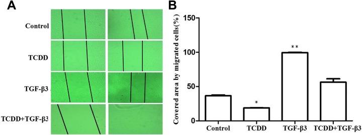
Figure. 3.
The effects of cell migration in MEPM cells. When MEPM cells were seeded and grew to confluence, the cells were scratched with a pipette tip, and 10 nM TCDD, 10 ng/mL TGF-β3, or combination of 10 nM TCDD and 10 ng/mL TGF-β3, or DMSO (≤0.05%) treated MEPM cells as the experiment group and the control group, respectively. After 24 hours treatment, the cells were photographed. The wound closure was determined using ImageJ software. Data were mean values ± standard deviation of 3 replicate experiments. *P < .05 or **P < .01 versus the corresponding control values. MEPM indicates mouse embryonic palatal mesenchymal; TGF-β3, transforming growth factor β3; TCDD, 2,3,7,8-tetrachlorodibenzo-p-dioxin.

