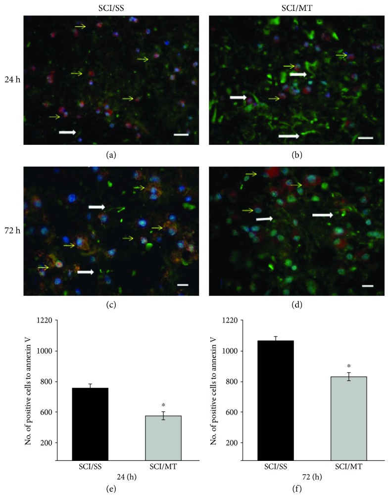
Figure 6.
Representative microphotographs of annexin V-positive cells (red, Alexa 546) and neurofilaments (green, Alexa 488) in the injury area after spinal cord contusion and sacrificed at 24 or 72 hours after damage. (a, c) Animals with spinal cord injury (SCI) plus saline solution (SS); (b, d) rats with SCI and treated with two doses of metallothionein (MT) starting at 2 and 8 h after the lesion; all animals were sacrificed at 24 h after SCI. (a, b) Animals with similar conditions described above for SS and (c) and SCI plus MT but sacrificed 72 hours after injury. Yellow arrows showed annexin V-reactive cells, as well as some immunoreactive fibers to NF (white arrows); the MT reduced neuronal damage and preserved a greater number of fibers, Scale bar 20 μm. (e, f): Number of annexin V-positive cells per mm3 measured at 24 or 72 h, respectively, after damage. The results are shown as average values ± SEM of 3–6 animals per group. Student's T test, ∗p < 0.05.

