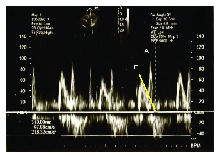
Figure 4.

Diagnosing diastolic dysfunction with echocardiography. Transmitral flow velocities measured with pulsatile wave Doppler technique. The ratio (E/A) of the early diastolic peak velocity (E) and the late diastolic velocity (A) is lower than 1. Deceleration time (DT) is the interval from the peak of the wave E to its end (marked with a yellow line). In this case, its prolongation was measured (310 sec). The above alterations prove the left ventricular diastolic dysfunction (relaxation disorder).
