Figure 1.
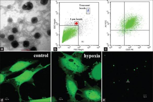
EMP formation and characterization. (a) TEM micrograph of EMPs released from HUVECs. (b) Fluorescent beads of 1 μm were used to define the MP gate (gate events <1 μm; MP gate: R1). (c) Double-positive events for Annexin V-FITC and CD31-PE were used to identify EMPs and count for each sample. (d) Confocal microscopic images of calcein-AM-labeled HUVECs released membrane blebbing and vesicles after hypoxia (right) in comparison with normoxia (left). (e) Confocal microscopic images of calcein-AM-labeled EMPs. MP: Microparticle; EMP: Endothelial MP; TEM: Transmission electron microscopy; HUVECs: Human umbilical vein endothelial cells.

