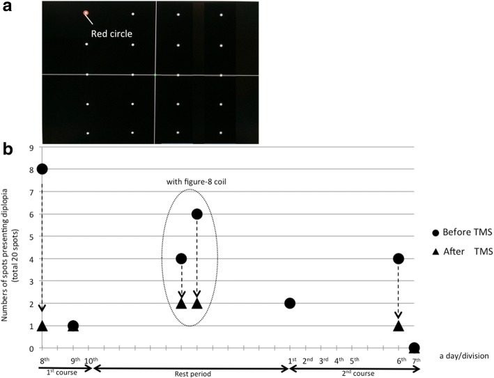
Fig. 2.
a: Initial arrangement of the spots used to detect locations presenting diplopia on a 20-in. PC monitor, b: Changes in the number of “diplopia-spots”. We started the assessment from immediately before and after the 8th TMS session, and thus did not assess all TMS sessions. We performed assessments in the 8th and 9th sessions in the 1st course and the 1st, 6th, 7th, 9thand 10th sessions in the 2nd course. The 8th, 9th and 10th sessions in the 2nd course are shown in Fig. 3. Dotted arrows show the immediate change in diplopia-spots with TMS. On the 7th intervention in the 2nd course, diplopia disappeared from the evaluation range, and therefore extended the range of assessment. On two days during the rest period between the 1st and 2nd courses, diplopia improved immediately after localized stimulation over the motor cortex with a Figure-8 coil (In a dotted oval)

