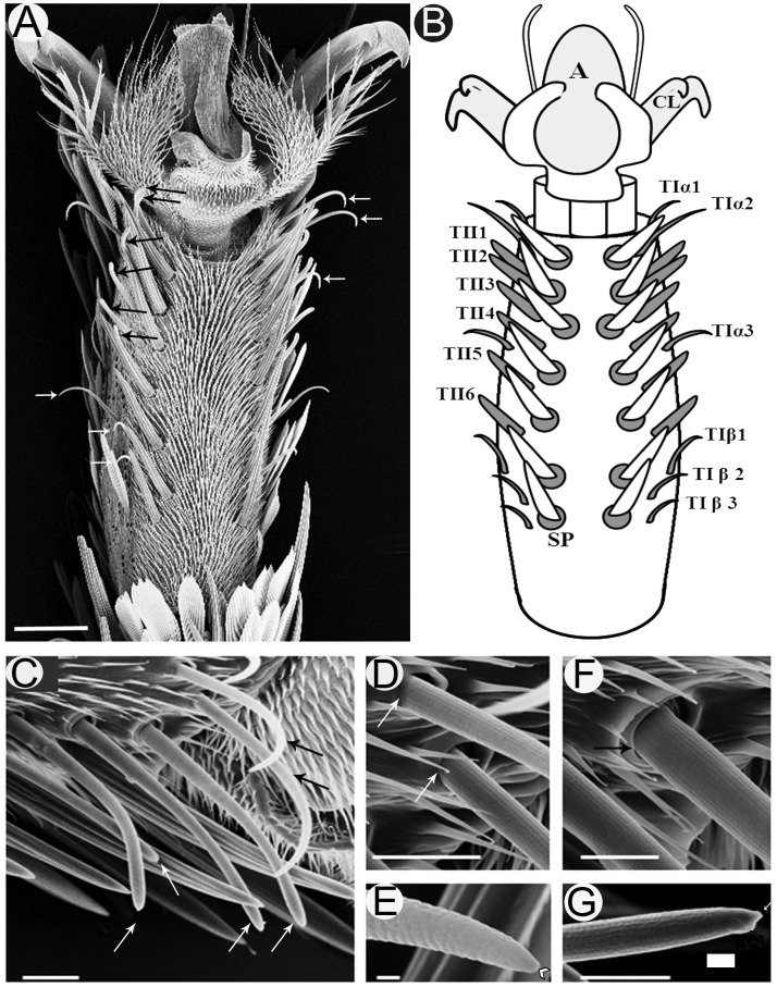
FIGURE 1.
Scanning electron micrographs of the ventral surface of the fifth tarsomere of female S. littoralis showing two parallel rows of sensilla chaetica. (A) These sensilla may be distinguished by their location and morphology as TIα (upper white arrows), TII (black arrows), and TIβ (lower white arrows) sensilla. (B) Schematic diagram illustrating the different sensillum types and numbers: TIα1-3, TII1-6, and TIβ1-3. (C) A magnified view of the anterior-lateral part of the fifth tarsomere showing the TIα1-2 (black arrows) and TII1-4 sensilla (white arrows). (D) The basal socket structure of the TI sensillum (white arrows), and (E) its fine tapering uniporous tip (black arrow). (F) The basal folded socket structure of the TII sensillum (black arrow), and (G) its blunt uniporous tip (white arrow). Notice the swollen knob close to the sensillum pore (arrow head). A, arolium; CL, claw; SP, spine. Scale bars: 50 μm in A; 20 μm in C; 1 μm in E; and 10 μm in D, F, and G.

