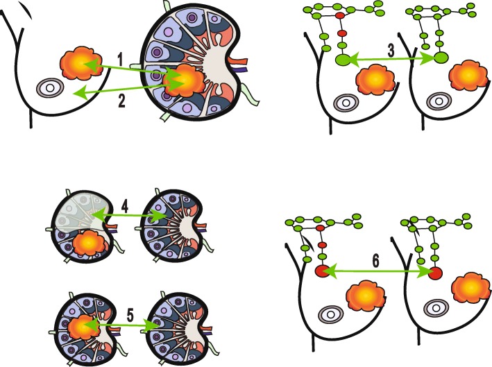
Fig. 2.
Different scenarios for studying lymph nodes, breast cancers and normal tissue. Six scenarios depict different comparisons (indicated by green arrows): scenario 1, involved lymph node versus primary tumour (number of studies = 8); scenario 2, involved lymph node versus normal breast tissue (number of studies = 1); scenario 3, uninvolved LNs in LN-positive patients versus uninvolved LNs in LN-negative patients (number of studies = 2); scenario 4, uninvolved residual portion of involved LN versus patient-matched uninvolved LN (number of studies = 2); scenario 5, involved LN versus patient-matched uninvolved LNs (number of studies = 1); scenario 6, involved sentinel LNs in patients with additional, non-sentinel, positive LNs versus involved sentinel LNs in patients with additional, non-sentinel, negative LNs (number of studies = 1). Tumours are shown in orange and red and green denote involved and uninvolved LNs, respectively. In scenario 4, the shaded portion represents the uninvolved residual portion of an involved LN

