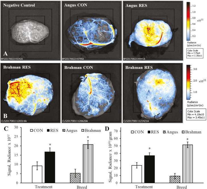
Figure 2.
Fluorescent detection of macroscopic cotyledonary blood vessel density in RES and CON-fed Angus and Brahman heifers at day 180 of pregnancy. Representative placentome images from Angus (A; top three images and scale bar) and Brahman heifers (B; middle three images and scale bar) with cotyledonary surface up. All treatment by breed interactions for macroscopic blood vessel density were P > 0.15. Main effects of dietary treatment and breed of absolute fluorescent signal intensity (C) and fluorescent signal intensity relative to individual placentome weights (D). Asterisk (*) represents a difference (P < 0.05) between dietary treatment groups or between breeds.

