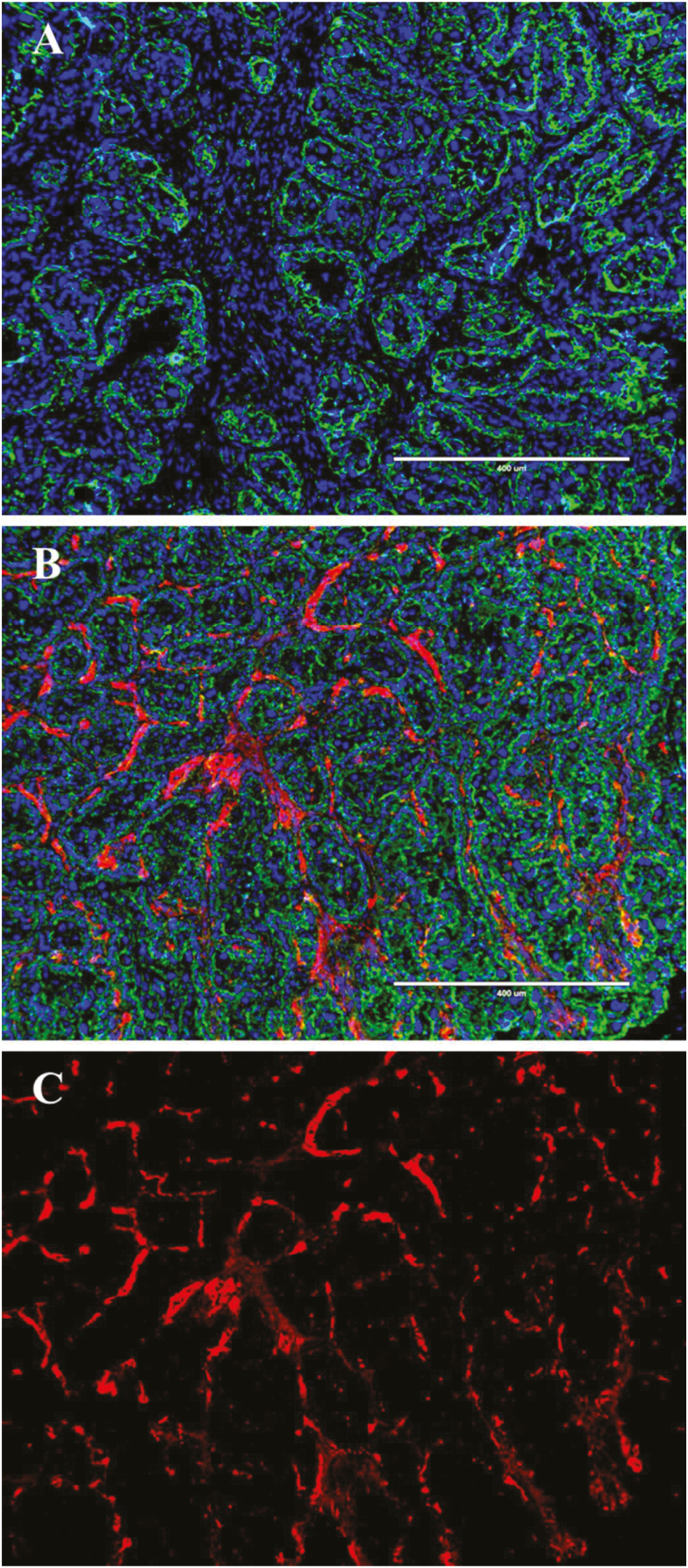
Figure 3.
Representative immunofluorescence images of heifer placentomes at 180 d of gestation. Cryosectioned placentomes were stained for capillaries with Anti-Von Willebrand Factor (red, Alexa Fluor 594), caruncular and chorionic epithelium (green, FITC), and nuclei (blue, DAPI). (A) represents a negative control–treated without Anti-Von Willebrand Factor. (B) represents a fully stained image with all 3 fluorescent channels overlaid and (C) represents the Texas Red channel only from (B). The white scale bar represents 400 μm.

