
Figure-3.
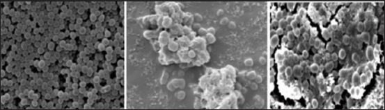
Dissolution of biofilm following UA treatment. It scanning electron microscopic image of non-biofilm Staphylococcus aureus (left). Biofilm-forming Staphylococcus aureus with intracellular adhesions and thick matrix (middle), and biofilm-forming S. aureus treated with UA showing decreased intracellular adhesions and thin matrix layer (right).