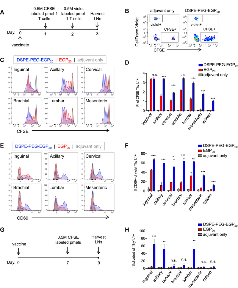
Figure 5: DSPE-PEG-peptides achieve broad lymphatic distribution and extended presentation.

A-F, C57BL6/J mice were immunized with 25 µg c-di-GMP and either 5 nmol EGP20 peptide, DSPE-PEG-EGP20 peptide, or no antigen. 24 hours (h) later, 0.5 million CFSE-labeled pmel-1 CD8+ T cells were transferred intravenously, and 48h after immunization, 0.5 million CellTrace Violet-labeled pmel-1 CD8+ T cells were transferred intravenously. LNs and spleens were isolated from recipient mice 24h later. A, Experimental timeline. B, Gating strategy to identify pmel-1 transferred populations, gated on live CD8+ T cells. C, Representative plots showing CFSE dilution among CD8+Thy1.1+ cells transferred 24h after immunization. D, Mean proliferation index of pmel-1 T cells in indicated LNs or spleen. n = 3/group; student’s t-test between EGP20 and DSPE-PEG-EGP20; representative of 3 independent experiments. E, Representative CD69 expression among CD8+Thy1.1+CellTrace violet+ cells transferred 48h after immunization. F, Mean %CD69+ of CD8+Thy1.1+CellTrace violet+ pmel-1 T cells in indicated LNs or spleen. n = 3/group; student’s t-test between EGP20 and DSPE-PEG-EGP20; representative of 3 independent experiments. G-H, Mice were immunized with 25 µg c-di-GMP and either 5 nmol EGP20 peptide, DSPE-PEG-EGP20 peptide, or no antigen. Seven days later, 0.5 million CFSE-labeled pmel-1 CD8+ T cells were transferred intravenously. LNs were isolated and processed 48h after transfer. F, Experimental timeline. G, % dividing CD8+Thy1.1+ in indicated LNs or spleen. n = 3/group; student’s t-test between EGP20 and DSPE-PEG-EGP20; representative of 2 independent experiments. *P<0.05, **P<0.01, ***P<0.001.
