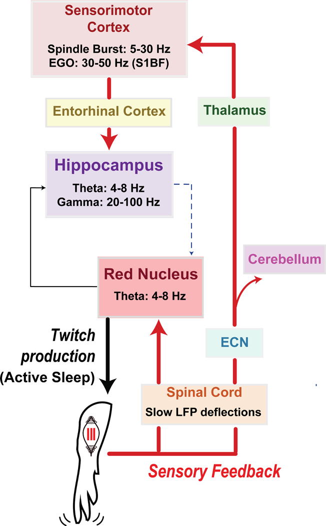
Figure 1. Diagram illustrating anatomical pathways conveying twitch-related sensory feedback in neonatal rats.

A twitch of the forelimb is generated in the red nucleus during active sleep. Sensory feedback (or reafference) from the twitch causes a cascade of precisely timed neural responses in sensorimotor structures across the neuraxis, including spinal cord, red nucleus, sensorimotor cortex, and hippocampus. Through repeated activation of these structures, twitches provide a unique opportunity for the synchronization of oscillatory activity. See text for further discussion.
