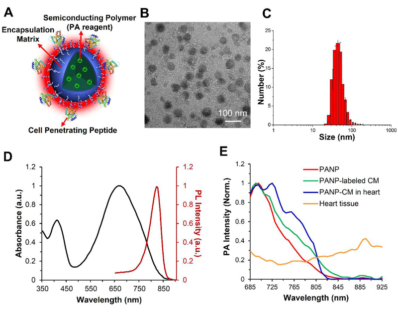
Figure 1. Characterization of semiconducting polymer (SP)-based photoacoustic nanoparticles (PANPs).
(A) The PANPs were composed of three components: SPs (Poly[2, 6-(4,4-bis-(2-ethylhexyl)-4H-cyclopenta[2,l-b;3,4-b’]dithiophene)-alt-4,7(2,l,3 benzothiadia-zole)]) as the PA contrast agent, polymer lipids as the encapsulant, and cell-penetrating peptides as the labeling enhancer. (B) The morphology was studied by transmission electron microscopy (TEM). (C) Particle size distribution of PANPs in water determined by a dynamic light scattering analyzer. (D) The maximum peaks of UV-visible absorption and photoluminescence spectrum for PANPs were at 670 nm and 820 nm, respectively. (E) Following multi-spectral excitations of near-infrared lasers (680–970 nm), pure PANPs showed a specific PA spectrum that reached a maximal peak at 705 nm and gradually decreased to zero after 850 nm (red). This narrow spectrum was consistent with PANP-labeled hESC-CMs (green), even when they were injected into the heart (blue). This spectrum had a largely different pattern and a much higher signal peak compared to the one from cardiac tissues (yellow).

