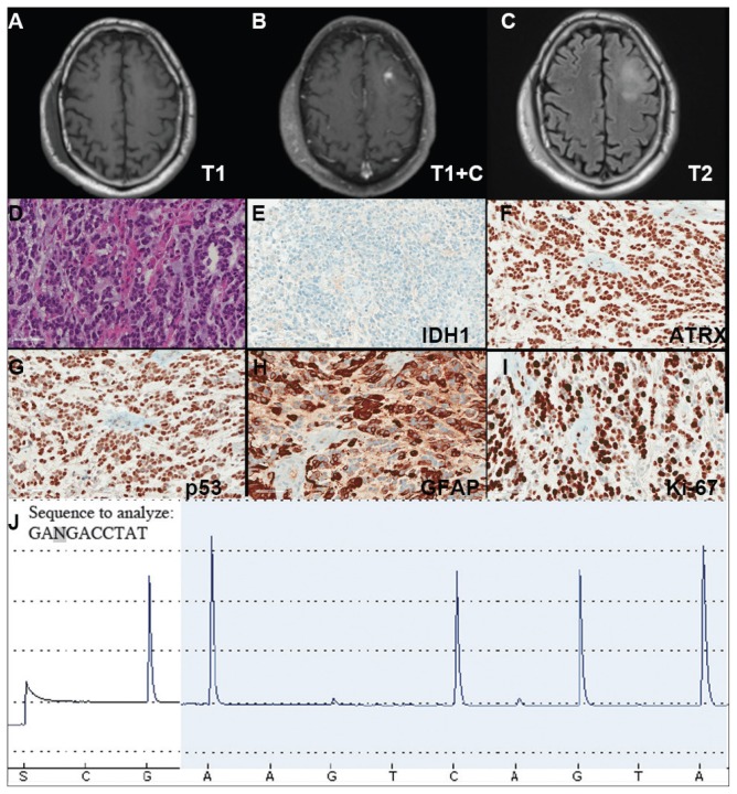
FIGURE 3. Case 3 Imaging, Immunohistochemical Stains, and Pyrosequencing.
(A–C) Imaging studies demonstrating a left frontal lesion; representative images of H&E hematoxylin & eosin (D) and indicated immunohistochemical (E–I) stained slides of lesion; (J) Pyrogram of IDH1 gene pyrosequencing (wild-type sequence GACGA corresponding to compliment of nucleotides 397–393 of IDH1 cDNA)

