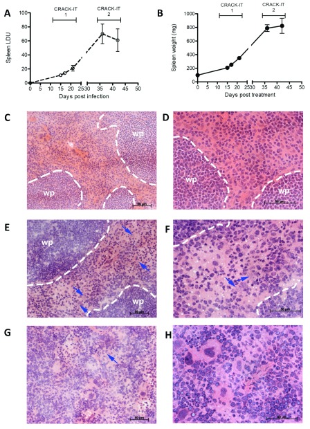
Figure 1. Splenic response to L. donovani infection.
Spleens from BALB/c mice infected with L. donovani were removed at 15-, 17-, 21-, 36- and 42-days post infection, along with spleens from naïve age matched controls. A. Parasite burdens were determined by impression smears and are represented as mean LDU ± SE (n=5 mice per time point). B. Spleen weight for naïve and infected mice (mg ± SE). C– H. Representative histology of spleens from naïve mice ( C and D) and from d17 ( E and F) and d42 ( G and H) infected mice. Hematoxylin and Eosin x20 ( C, E and G) and x40 ( D, F and H); scale bars 50 µm. Blue arrows highlight parasite clusters. White dashed lines indicate red pulp (rp) / white pulp (wp) boundary. G and H show increased frequency of megakaryocytes, indicative of extramedullary haematopoiesis and loss of white pulp / red pulp discrimination. Data are pooled from two independent experiments covering the early and late phase of infection.

