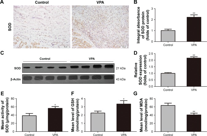
Figure 4.
VPA inhibited oxidative stress in random skin flaps.
Notes: (A) SOD expression in each group as assessed by IHC (original magnification ×200). (B) The optical density values of SOD. (C) Protein expression of SOD in each group as assessed by Western blot analysis. The gels have been run under the same experimental conditions, and cropped blots are used here. (D) Densitometry result of SOD protein expression in the two groups. (E–G) The levels of SOD, GSH, and MDA in random skin flaps were determined using the xanthine oxidase method, 5,5′-dithiobis method, and thiobarbituric acid test, respectively. Values are expressed as mean ± SEM, n=6 per group. *P<0.05 and **P<0.01, vs control group.
Abbreviations: GSH, glutathione; IHC, immunohistochemistry; MDA, malondialdehyde; SEM, standard error of mean; SOD, superoxide dismutase; VPA, valproic acid.

