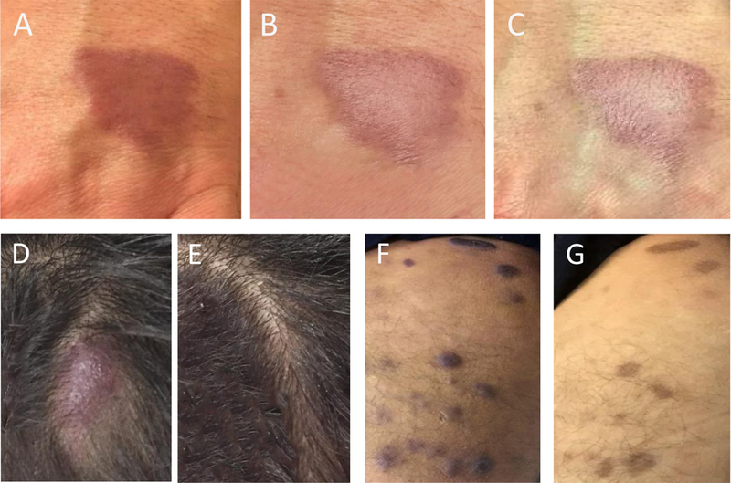
Figure 1. Baseline and post-therapy lesion photographs.

(A-C) Case example (patient 2). Left hand lesion (A) pre-therapy, (B) post 4 weeks and (C) post 8 weeks of therapy. (D-G) Case example (patient 9). Scalp lesion (D) pre-therapy and (E) post 6 weeks of therapy. Right medial thigh lesion (F) pre-therapy and (G) post 6 weeks of therapy.
