Figure 4.
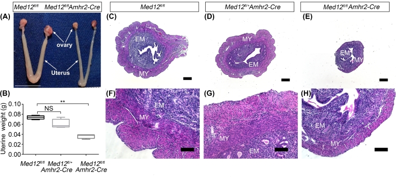
Med12fl/fl Amhr2-Cre uterine anatomy and histology. (A) Gross morphology of 12-week-old female mouse reproductive tract. The Med12fl/fl (control mice) display normal reproductive tract. In comparison to the controls, Med12fl/fl Amhr2-Cre reproductive tract is atrophic. Scale bar = 1cm. (B) Twelve-week-old uterine weight is significantly decreased in Med12fl/fl Amhr2-Cre as compared to Med12fl/fl and Med12fl/+ Amhr2-Cre group (n = 7). Data are presented as mean ± SEM. **P < 0.01. (C–H) Hematoxylin and eosin staining of uteri from 12-week-old Med12fl/fl, Med12fl/+ Amhr2-Cre and Med12fl/fl Amhr2-Cre mice synchronized with PMSG and hCG. Note the atrophic uteri of Med12fl/fl Amhr2-Cre female. EM, endometrium; MY, myometrium. Scale bars = 100 μm.

