Figure 8.
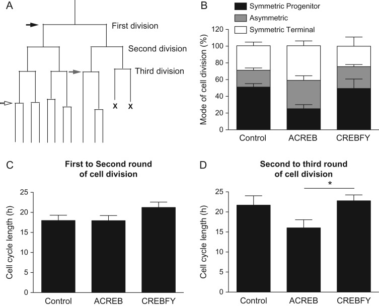
CREB signaling does not affect mode of cell division and cell cycle of neural cortical progenitors. (A) Schematic lineage tree indicating the mode of cell division (black arrow: SP—both daughter cells undergo new cycle of cell division; gray arrow: asymmetric—one daughter cell undergo new cycle of cell division and the other becomes postmitotic; white arrow: ST—both daughter cells become postmitotic) and rounds of cells division (first, second, and third division). “X” indicates cell death. (B) Mode of cell division of individual progenitor cells after transfection with GFP (control), A-CREB-GFP, or CREBFY-GFP (see also Supplementary Fig. 5 and Supplementary Movies 3 and 4). (C–D) Cell cycle length of individual progenitor cells followed by time-lapse video microscopy. Data show the time in hours between first and second rounds of cell divisions (C) or between second and third rounds cell divisions (D). Note that only in the later A-CREB-GFP expression shortens the cell cycle length as compared with CREBFY-GFP (One-Way ANOVA followed by Tukey's post hoc test; *P < 0.05). Number of trees analyzed—Control: n = 310; A-CREB: n = 106; CREBFY: n = 140.

