Fig. 4.
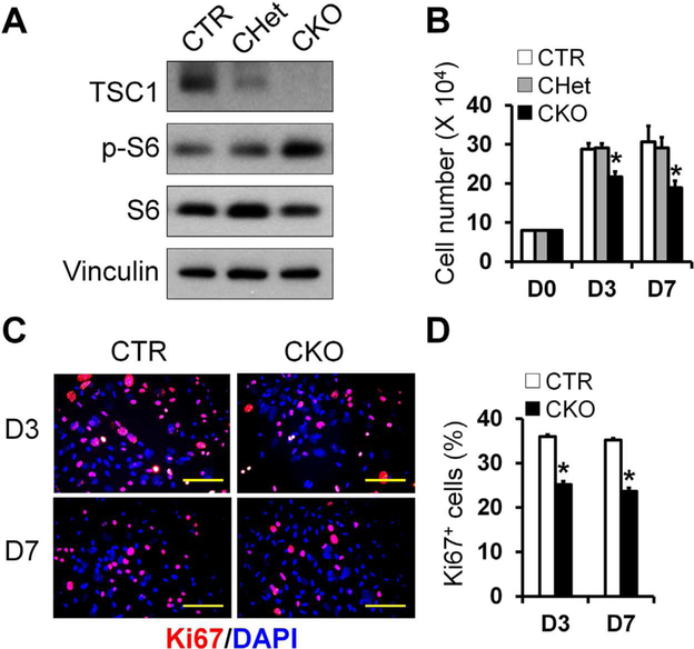
TSC1-deficient BMSCs have decreased proliferation. BMSCs were isolated from femur and tibia of CTR, CHet, and CKO mice as described in Materials and Methods. (A) TSC1 was efficiently deleted in BMSCs in association with increased phosphor-S6 level in CKO group as shown by Western blot analysis. (B) Quantitation of cell number cultured for 3 and 7 days in osteogenic medium. n = 3 per group. (C) Representative merged fluorescent images of Ki67 (red) and DAPI (blue) staining in CTR and CKO osteoblasts at 3rd and 7th day, in the same cultures system as B. Scale bar = 50 μm. (D) Quantification of Ki67-positive (Ki67+) in the cultures shown in C. n = 3 per group, *p < 0.05. Values are presented as mean + SE. DAPI = 4,6-diamidino-2-phenylindole; CTR = control (Tsc1F/F).

