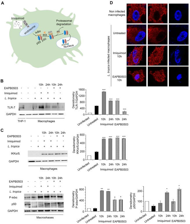
Fig 2. Imiquimod triggers an increase of TLR-7 expression in L. tropica infected macrophages leading to the canonical NF-κB pathway activation.
Schematic representation of the mode of action of Imiquimod through TLR-7 immuno-modulation and its subsequent downstream NF-κB activation (A). Western blot analysis for TLR-7 (B), IKKα/β, P-IκBα and P50 (C) in L. tropica infected macrophages treated with 0.1 μM of Imiquimod or EAPB0503 for 10 and 24h. The results depict one representative experiment among three independent ones. Densitometry was performed using Image Lab software (Biorad). Results shown represent the average of quantification of three independent experiments. (D) Confocal microscopy on L. tropica infected macrophages treated with 0.1 μM of Imiquimod or EAPB0503 for 10h. The NF-κB p65 subunit was stained with an anti-p65 antibody (red), and nuclei were stained with Hoechst 33342 (blue). The results depict one representative experiment among three independent ones.

