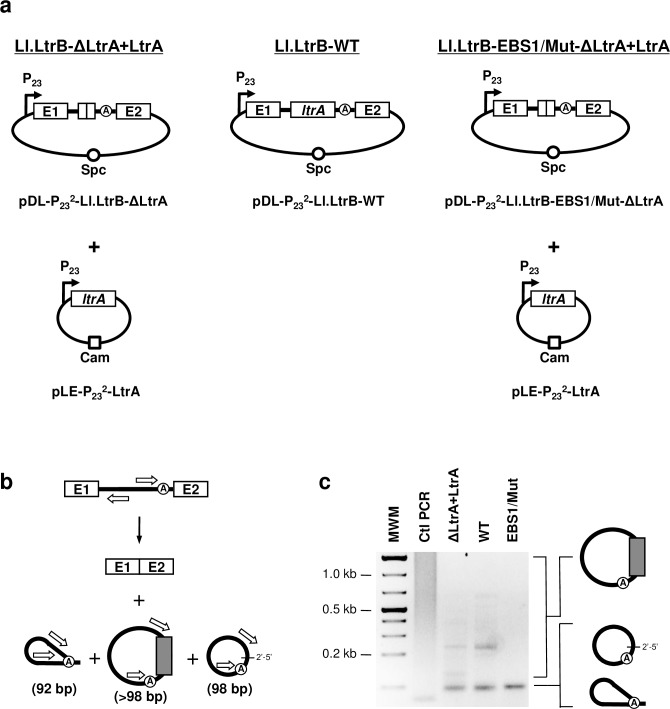
Fig 2. Detection of mRNA fragments at the splice junction of excised intron RNA circles.
(a) Various Ll.LtrB constructs used in this study where the LtrA protein is provided either in trans (Ll.LtrB-ΔLtrA+LtrA, Ll.LtrB-EBS1/Mut-ΔLtrA+LtrA) or in cis (Ll.LtrB-WT) (b) Schematic of Ll.LtrB self-splicing. Position of the primers (open arrows)(S2 Table) used to amplify the splice junction of excised introns by RT-PCR is depicted (92 bp (lariat), 98 bp (circle) or >98 bp (circle harboring additional nts)). (c) RT-PCR amplifications of intron splice junctions. Amplifications were performed on total RNA extracts from L. lactis (NZ9800ΔltrB) harboring different Ll.LtrB constructs expressed under the control of the P23 constitutive promoter (ΔLtrA+LtrA: pDL-P232-Ll.LtrB-ΔLtrA and pLE-P232-LtrA)(WT: pDL-P232-Ll.LtrB-WT)(EBS1/Mut: pDL-P232-Ll.LtrB-EBS1/Mut-ΔLtrA and pLE-P232-LtrA). The EBS1/Mut intron variant was shown to splice accurately and efficiently in vivo by RT-PCR amplifications of both released introns and ligated exons. Additional nts incorporated at the junction of intron circles are represented by a gray box.

