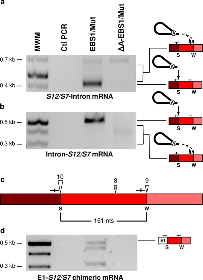
Fig 8. Detection of intermediates unique to the reverse splicing pathway: Ll.LtrB reverse-spliced within an L. lactis mRNA and an E1-mRNA chimera.
RT-PCR assays were performed to detect the 5’ and 3’ junctions of Ll.LtrB-EBS1/Mut-ΔLtrA+LtrA and Ll.LtrB-ΔA-EBS1/Mut-ΔLtrA+LtrA reverse splicing events within the Ribosomal Protein S12/S7 mRNA (a, b). Complete and dashed arrows indicate reverse splicing of Ll.LtrB lariats within strong (S) and weak (W) IBS1/2-like sequences (—|—) respectively. The strong (S)(10/11 nts)(large arrowhead) and weak (W)(8-9/11 nts)(small arrowheads) IBS1/2-like sequences invaded by reverse splicing are represented (c). The sites flanking the mRNA fragment (c, 161 nts) initially detected at intron circle splice junctions (Fig 3B) are indicated by open arrowheads. mRNA chimeras between ltrB-exon 1 (E1) and L. lactis mRNAs (d, E1-S12/S7) were also detected by RT-PCR at IBS1/2-like sequences.

