Figure 2.
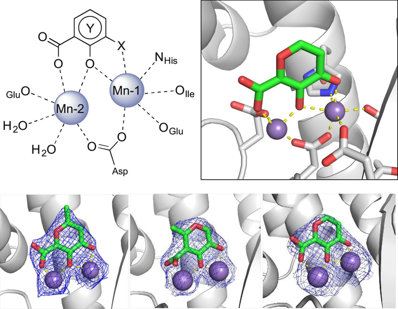
Top left: Representative chemotype shared by active MBP fragments of influenza PAN highlighting the coordination motif of these MBPs to the dinuclear active site metal ions. Top right: X-ray co-crystal structure of compound 3 (PDB ID: 6DCZ) in the PAN active site. Mn-2 is also coordinated by two water molecules at the open coordination sites (not shown). Bottom: X-ray co-crystal structure of compounds 1 (left; PDB ID: 6DZQ), 2 (middle; PDB ID: 6DCY), and 3 (right; PDB ID: 6DCZ) in the PAN active site. All ligands were found to coordinate in a similar manner. Compound 2 coordinates Mn-2 with the electron density (blue mesh is the 2FoFc map contoured to 2σ) corresponding to the carboxylic acid moiety found to be diffuse above the metal center. The poorer coordination ability of compound 2 is likely due to internal steric pressure exerted by the methyl group residing alpha to the carboxylic acid.

