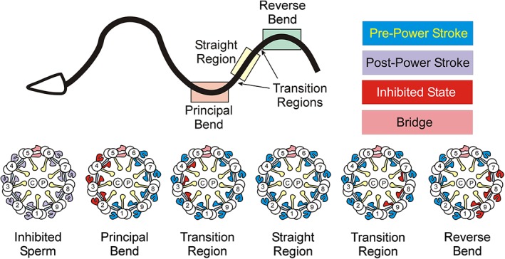
Figure 3.

Dynein power stroke transitions during oscillatory beating. Top – Diagram of a swimming sea urchin sperm. The symmetric flagella waveform propagates from base to tip. Regions corresponding to the principal bend (light pink), reverse bend (light green), and the straight region (light yellow) between them are boxed. Also shown (arrows) are the “principal bend to straight region” and “straight region to reverse bend” transition segments where only some inner arms are in the inactive conformation. Bottom – Diagrams of axonemal cross‐sections showing the status of individual dynein motors on each doublet in EHNA‐inhibited sperm, principal and reverse bends, straight intervening region and the transitional segments; blue – pre‐power stroke, purple – post‐power stroke, red – inhibited (weakly bound or microtubule‐detached) state, pink – bridge structure between doublets 5 and 6, blue‐gray – N‐DRC, yellow – radial spokes. In bends, both inner and outer arms on doublets 2–4 or 7–9 are in the inhibited state. In the straight region, most dyneins are in a pre‐power stroke conformation, while in transitions between bends and straight segments, only inner arm subsets are inactivated. EHNA, erytho‐9‐(2‐hydroxynonyl)adenine; N‐DRC, nexin‐dynein regulatory complex. [Color figure can be viewed at http://wileyonlinelibrary.com]
