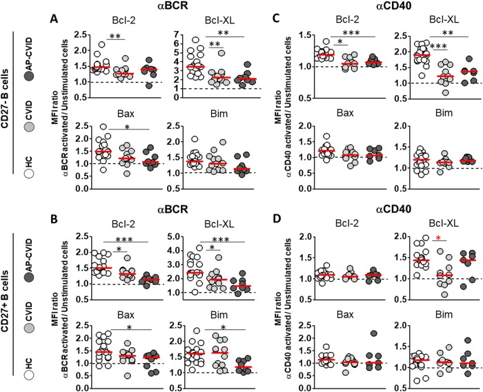
Fig. 5. Heterogeneous stimulation-induced expression of Bcl-2 and Bcl-XL between CVID patients after naive and memory B cell activation.
MFI ratios of Bcl-2 (upper left), Bcl-XL (upper right), Bax (lower left) and Bim (lower right) in naive CD19+CD27− and memory CD19+CD27+ B cells, unstimulated, and after activation with anti-BCR (a, b) or anti-CD40 (c, d) in healthy controls (white circles), CVID (light gray circles), and apoptosis-prone CVID (dark gray circles) patients. Data are given as medians (Kruskal–Wallis test P values: *P < 0.05*; **P < 0.01; ***P < 0.001). MFI median fluorescence intensity, HC healthy controls (n = 20), CVID common variable immunodeficiency patients (n = 12), AP-CVID apoptosis-prone CVID patients (n = 8)

