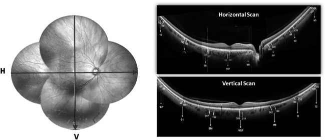
Figure 4.
Shows a representative fundus photograph (montage) of right eye with arrows showing the direction of line scans across both horizontal and vertical meridian (left side). Top-right and top-bottom shows representative wide-field optical coherence tomography (WF-OCT) in horizontal and vertical meridian respectively showing the choroidal thickness (CT) and large vessel choroidal thickness (LCVT) in macular segments and all four quadrants. HSF, horizontal subfoveal; VSF, vertical subfoveal; TM, NM, SM, IM represent temporal, nasal, superior and inferior macular points respectively. T1, T2, N1, N2, N3, S1, S2, I1 and I2 are points of measurements in respective quadrants.

