Figure 5.
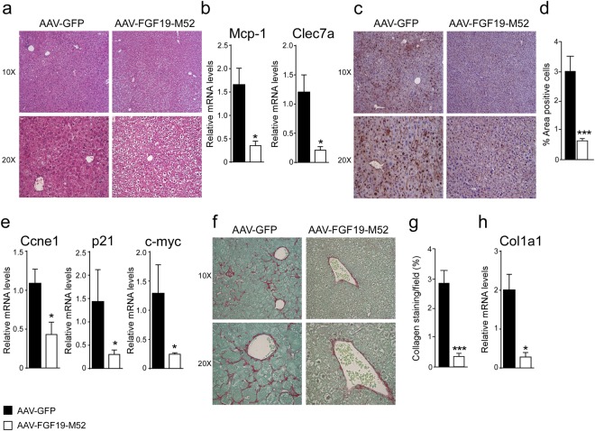
FGF19-M52 analogue protects Fxr−/− mice from fibrosis and cellular proliferation. (a) Liver histology assessed by hematoxylin and eosin (H&E) staining and observed by light microscopy (magnification, 10–20X). (b) Mcp-1 and Clec7a gene expression levels assessed by real time qPCR in tumor-free liver extracts. (c) Hepatic immunohistochemical staining of the oncogene Ccnd1. (d) Quantification of Ccnd1 staining assessed with ImageJ and expressed as % of the area occupied by positive cells. (e) Ccne1, p21 and c-myc gene expression levels assessed by real time qPCR in tumor-free liver extracts. Expression was normalized to Cyclophillin. (f) Hepatic collagen deposition assessed by Sirius Red staining (magnification, 10–20X). (g) Quantification of collagen deposition assessed with ImageJ and expressed as % of collagen staining/field (h) Col1a1 gene expression levels assessed by real time qPCR in tumor-free liver extracts. Expression was normalized to Cyclophillin. All values represent mean ± SEM. Statistical significance (**p < 0.01, ***p < 0.001) was assessed by Mann-Whitney’s U test.

