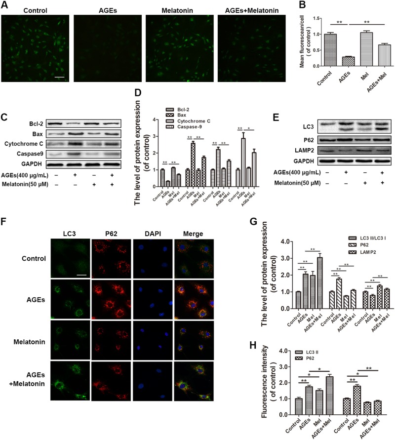
Fig. 2. Melatonin reduces mitochondrial functional damage and promotes autophagy flux in AGE-treated EPCs.
EPCs were pre-treated with 50 μM melatonin for 2 h and then 400 μg/mL AGEs were added for an additional 24 h. a, b Mitochondrial permeability transition pore (mPTP) opening was detected by co-loading with calcein-AM and cobalt. Scale bar, 50 μm. c, d Protein expression levels of Bcl-2, Bax, cytochrome c, and caspase-9 in EPCs of each group treated as described above. e, g Protein expression levels of LC3, p62, and LAMP2 in EPCs treated as described above. f, h Double immunofluorescence of LC3 protein (green) and p62 protein (red) in EPCs. Scale bar, 25 μm. Data are presented as the mean ± SEM. Significant differences between the treatment and control groups are indicated as **P < 0.01 or *P < 0.05. n = 3

