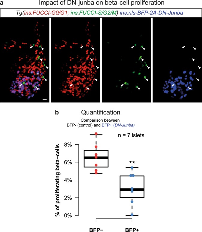
Figure 3.
Inhibition of junba reduces the proliferation of zebrafish beta-cells. (a) Maximum intensity confocal projections of beta-cells from a 30 dpf animal showing mosaic expression of nls-BFP-2A-DN-junba (blue) together with Tg(ins:FUCCI-S/G2/M) (green) and Tg(ins:FUCCI-G0/G1) (red). Arrowheads mark proliferating beta-cells, as indicated by the presence of green fluorescence and absence of red fluorescence. Scale bar 10 μm. (b) Tukey-style boxplots showing the percentage of proliferating beta-cells among BFP+ and BFP- cells. BFP+ cells co-express DN-junba, while the BFP- cells act as internal control. The BFP+ cells show a significant decrease in the proportion of proliferating cells (t-test, **p-value < 0.01). ‘n’ denotes number of islets.

