Fig. 1.

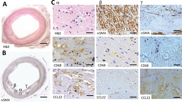
Expression and localization of CCL22 in a human common carotid artery
Fig. A and Fig. B are low-power magnification images of H&E (H & E) and α-smooth muscle actin (αSMA) stains. In Fig. B, α signifies an area of intermediate vascular smooth muscle (VSMC) cell density and the accumulation of foamy macrophages, β signifies an area with a high degree of VSMC density and γ signifies an area with virtually no VSMC. Figures C-α are high-power magnified images showing H & E and immunohistochemical stains for CD68 and CCL22, respectively, of the α-area in Fig. B. Fig. C-β shows immunohistochemical staining for αSMA, CD68, and CCL22, respectively, of the β-area in Fig. B. Fig. C-γ shows immunohistochemical staining for αSMA, CD68, and CCL22 of the γ-area in Fig. B. At first glance, the distribution of VSMCs appeared heterogeneous, even in atherosclerotic lesions that were concentric (A, B).
CD68 positive foamy macrophages were also positive for CCL22 (C-α). Macrophages in close proximity to VSMC densities were CCL22 negative (C-β). VSMCs were not present around cholesterol clefts; however, positive CCL22 staining was present (C-γ). Bars indicate 1000 µm (A and B), 10 µm (C-α), 100 µm (C-β αSMA and C-γ αSMA), and 50 µm (C-β and C-γ, CD68 and CCL22). The negative control staining of Fig. C-γ CCL22 was shown in Supplemental Fig. 1A.
