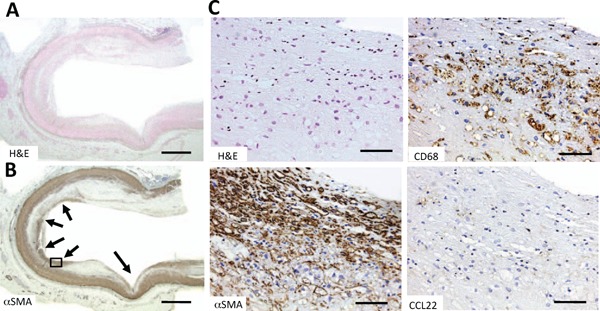
Fig. 2.

Histochemical staining of CCL22 in a human coronary artery with a bare metal stent
Fig. A and Fig. B are low-power magnified images of H&E (H & E) and α-smooth muscle actin (αSMA) stains, respectively. Arrows in Fig. B indicate the former positions of stent-struts. Fig. C shows high-power magnified images of H & E, αSMA, CD68, and CCL22 stains, respectively, of the area in the box in Fig. B. Densities of VSMCs were high in the intima of the former contact sites of the stent-struts (Fig. C αSMA) while macrophages at these sites were CCL22-negative (Fig. C CCL22).
Bars indicate 1000 µm (Fig. A and B) and 100 µm (Fig. C).
