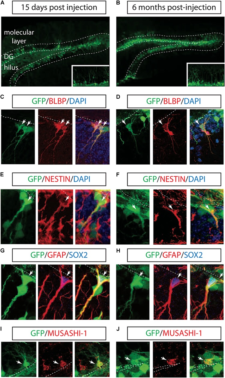
FIGURE 1.

Long-term marking of hippocampal NSCs by LV PGK-GFP. LV PGK-GFP was unilaterally injected into the hippocampal DG; brain sections were analyzed 15 days (A) and 6 months (B) later. GFP expression was evident in the DG at both time points. A higher magnification view is displayed in insets (A,B). GFP-expressing cells co-labeled with NSCs markers such as BLBP (C,D), NESTIN (E,F), SOX2, GFAP (G,H), and MUSASHI-1 (I,J) (arrows). Note that some GFP-positive cells stained for SOX2 showed co-localization with radial glial cell markers such as GFAP in their processes (G,H). DG, dentate gyrus; SGZ is marked with dotted lines.
