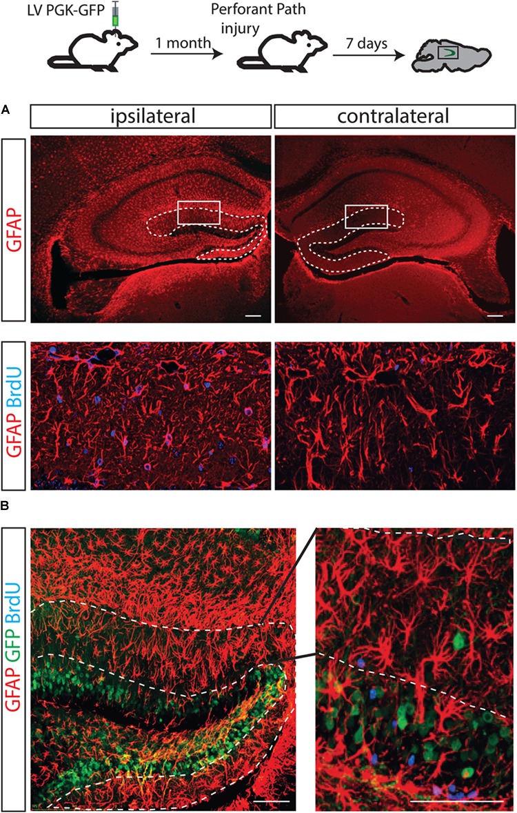
FIGURE 4.

The fate of hippocampal NSCs did not change following PP injury. A brain lesion in the perforant path resulted in over-expression of GFAP-positive cells in the molecular layer of the injury side but not the contralateral side (A, dotted line). As expected, an increased number of astrocytes (positively stained with BrdU) was found in the molecular layer of the ipsilateral side (B, left) but not in the contralateral side of these animals (B, right); however, GFP-labeled progenitors did not contribute to the increased astrocyte numbers.
