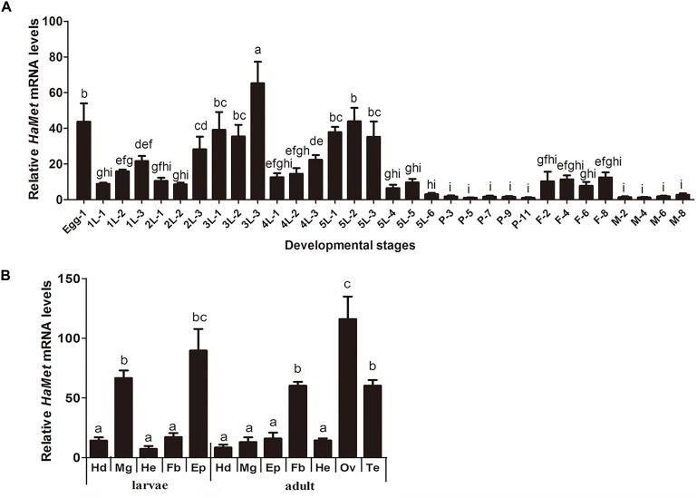
FIGURE 2.
Tissue and temporal expression patterns of HaMet. (A) Temporal expression of HaMet at different developmental stages. The samples are prepared at an interval of 1 day during larval stages and 2 days for pupae and adult. (B) Tissue distribution of HaMet transcripts in adults and 5th-instar larvae. Hd, head; Mg, midgut; He, head; Fb, fat body; Ep, epidermis; Ov, ovary; Te, testis. The bars represent the average (±SE) of biological repeats. Different letters indicate significant difference between specimens (P < 0.05).

