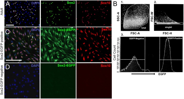
Figure 4.
Culturing spiral ganglion glial cells harvested from adult CD1 mice and sorted from neonatal Sox2-EGFP mice. (A) Spiral ganglion glial cells harvested from adult CD1 tissue cultured for 3 weeks and re-plated onto 2% Matrigel coated dishes. Glial cells (Sox2, green; Sox10, red) are observed to culture along with other non-neuronal, non-glial cells. Scale bar; 100 μm (B) Spiral ganglion single cell suspensions harvested from one-day old Sox2-EGFP mice were analyzed for the expression of EGFP. Representative fluorescence-activated cell-sorting analysis gating strategy is shown. First cells were gated for (1) forward and side scatter of dissociated cells followed by (2) gating for doublet discrimination, and lastly for (3) EGFP expression. A representative histogram with gating of EGFP+ and EGFP- populations is shown. (C,D) Spiral ganglion glial cells harvested from P0 Sox2-EGFP tissue and sorted into Sox2-positive and Sox2-negative fractions using a fluorescence activated cell sorter based on EGFP intensity. Glial cells nearly exclusively make up the positive sorted fraction (B) as based on Sox2-EGFP and Sox10 expression. No glia were observed in the Sox2-negative fraction. Scale bar; 10 μm.

