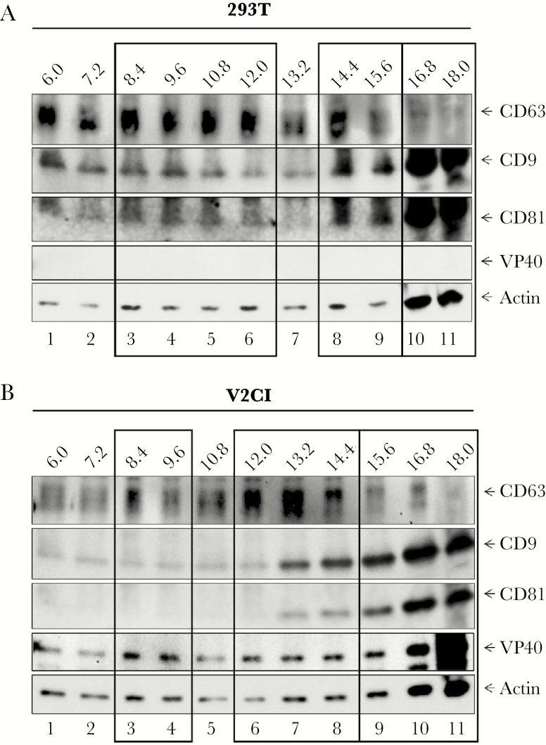
Figure 6.
Iodixanol gradient separation of extracellular vesicles (EVs) from 293T and VP40-producing cells. 293T and V2CI cells were grown in exosome-free media for 5 days, followed by harvesting of the supernatant and incubation with ExoMAX (1:1 reagent/filtered supernatant) reagent overnight at 4°C. The EVs were pelleted, resuspended in 300 µL of sterile 1× PBS, and loaded onto a 6–18% iodixanol density gradient (1.2% increments). Samples were ultracentrifuged for 90 minutes at 100000 ×g, followed by harvesting and isolation of each fraction, and incubation with 30 µL of NT80/82 particles overnight at 4°C. The NT pellets were washed in 1× PBS, resuspended in 12 µL of Laemmli buffer, and loaded onto a 4–20% Tris-glycine gel. Western blot of 293T (A) and V2CI (B) fractions were analyzed for levels of VP40, CD63, CD81, CD9, and Actin. Major groups of EVs or exosome type are indicated by black boxes.

