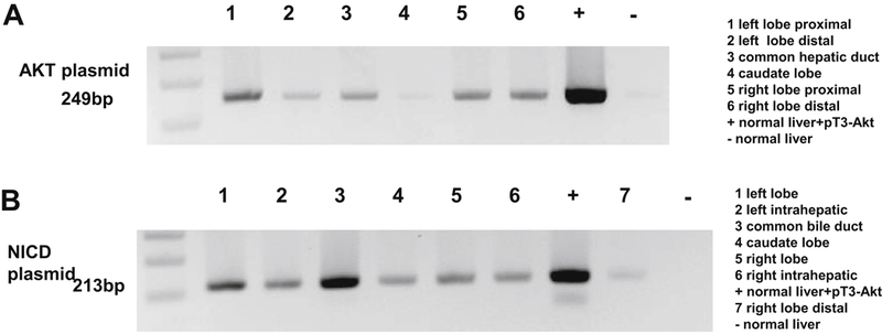
Figure 2: PCR analysis performed on genomic DNA collected from swine by amplification with primers for the human AKT and NCID gene in swine liver and bile duct.

A: Plasmid DNA AKT presence in liver tissue 21 days after ERCP and hydrodynamic injection. At day 21, swine liver were harvested, and DNA was extracted. PCR analysis of 6 samples from each swine liver was performed on genomic DNA by 35 cycles of amplification, denature at 94°C denature, anneal at 59°C anneal with primer for human AKT gene. B: Plasmid DNA NICD presence in liver tissue 30 days after ERCP and hydrodynamic injection. At day 30, swine liver were harvested, and DNA was extracted. PCR analysis of 6 samples from each swine liver was performed on genomic DNA by 35 cycles of amplification, denature at 94°C denature, anneal at 63°C anneal with primer for human NICD gene. Results show that each location has the plasmid AKT and NICD sequence expression.
