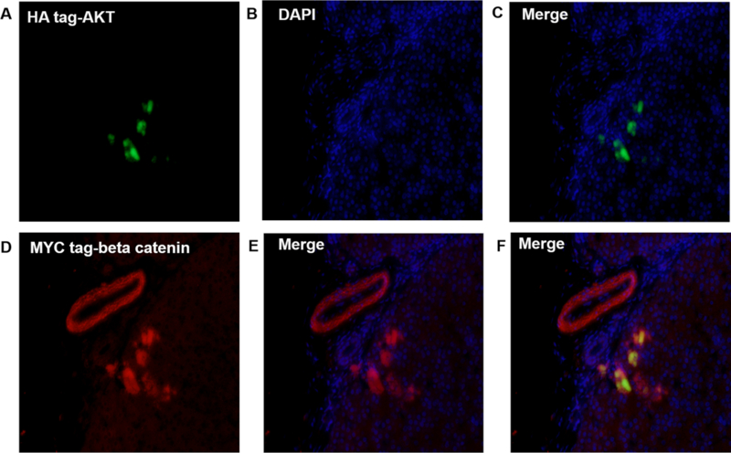
Figure 5: HA-Tag AKT and MYC-tag beta catenin protein were expressed in swine hepatocytes and bile duct.

Swine liver tissue that underwent hydrodynamic injection with AKT and beta-catenin plasmids were harvested at day 60, and tissues were analyzed for the expression of plasmid AKT and beta catenin protein via fluorescence microscopy. A–C show the anti HA-tag AKT protein inside hepatocytes. D–E demonstrates beta-catenin expressed in bile duct and hepatocytes. F illustrates that some hepatocytes express both AKT and beta-catenin. The figure demonstrate that hydrodynamic injection of plasmids can be stably integrated and expressed in swine hepatocytes and bile duct (amplification: 20X).
