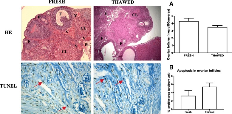
Fig. 2.
Photomicrographs of fresh and thawed ovarian tissue by HE staining and immunohistochemistry for apoptosis by TUNEL. Morphology is similar between groups. a Quantification of viable ovarian follicles in a 500-μm2 area, data are shown as the mean ± SD. Immunohistochemistry was performed on the ovarian follicles; dark brown-stained cells are considered positive (red arrow). b Results are expressed as a percentage of the positive area (arbitrary unity/mm2). Non-significant differences between groups in both analyses. p > 0.05, paired t test. HE × 100. TUNEL × 400. F follicles, CL corpora lutea, Fi fibrosis, V vessel

