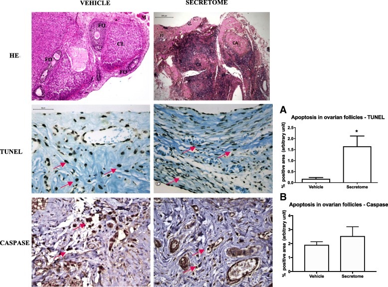
Fig. 4.
Photomicrography of cryopreserved ovarian grafts treated with vehicle or ASC secretome 30 days after an autologous avascular transplant. Treatment with secretome induced atrophy of the graft, with predominance of fibrosis and few viable follicles (HE). Immunohistochemistry for apoptosis (TUNEL and cleaved-caspase-3) was performed on the ovarian follicles, and dark brown-stained cells were considered positive (red arrows). The results are expressed as a percentage of the positive area (arbitrary unity/mm2). Apoptosis increased in TUNEL assay in grafts treated with the secretome (a) and remained similar in caspase (b). *p < 0.05, unpaired t test. HE × 50. Immunohistochemistry × 400. ASC adipose tissue-derived stem cells, CL corpora lutea, CA corpora albicans, FI fibrosis

