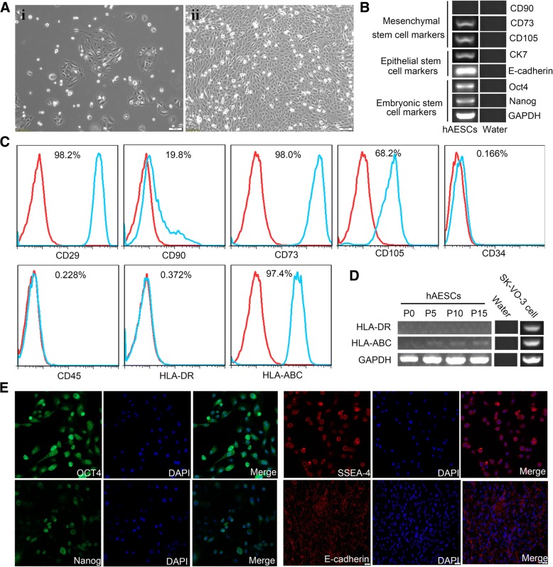
Fig. 1.
Characteristics of cellular morphology and expressions of markers in hAESCs. a Phase-contrast microscopic images of cultured hAESCs without (i) or with EGF (10 ng/ml) (ii). b RT-PCR analysis for the expressions of markers in hAESCs. Human AESCs specifically expressed various markers of CD73, CD105, CK7, E-cadherin, Oct4, and Nanog, but not CD90. Water was used as negative control. c Detection of surface markers in hAESCs (blue) and in isotype controls (red) by flow cytometry. Human AESCs expressed CD29, CD73, CD105, and HLA-ABC, but not CD90, CD34, CD45, and HLA-DR. d RT-PCR analysis for expressions of the major histocompatibility proteins. There were no expression of HLA-DR or low expression of HLA-ABC in hAESCs during passages 0 to 15. Water was used as negative control and ovarian cancer cell SK-VO-3 were used as positive control. e Immunofluorescence staining for the markers. All of hAESCs expressed the antigens of Oct4, SSEA-4, Nanog, and E-cadherin

