Fig. 3.
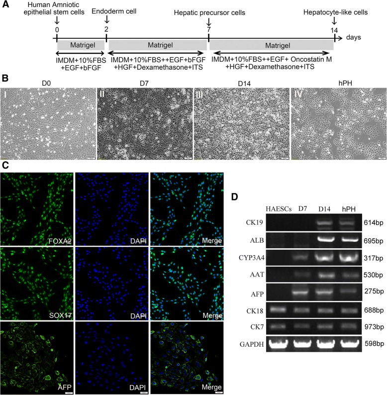
Differentiation of hAESCs into hepatocyte-like cells. a Schematic diagram of hAESCs differentiation into hepatocyte-like cells. b Images of the sequential morphological changes from hAESCs to hepatocyte-like cells. (I) Morphology at the preinduction stage of hAESCs. (II) The cell morphology has become polygonal in shape after 7-day hepatic lineage commitment medium induction. (III) At day 14, the cell morphology had become cuboidal in shape. (IV) The morphology of hPH. c Immunocytochemical examination of endoderm cells and hepatic precursor cells. The vast majority of induced cells expressed the definitive endoderm marker FOXA2 and SOX17 at day 2 and AFP at day 7 after induction of the differentiation procedure. d Expressions of the hepatocyte markers in the various stages of differentiation. After 0, 7, and 14 days of induction, the expressions of hepatocyte markers CK7, CK18, AFP, AAT, CYP3A4, ALB, and CK19 of hAESCs differentiated cells and hPH were analyzed by RT-PCR

