Fig. 6.
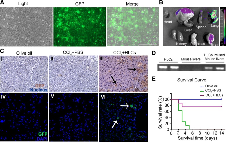
The distribution and therapeutic effects of HLCs in lethal acute liver failure mice. a Representative images of GFP-expressing HLCs. b The distribution of GFP marked HLCs in vivo of acute liver failure mice on day 3 after transplantation. HLCs mainly distributed in the lung and liver. c Immunohistochemical staining and immunostaining analysis of GFP-expressing HLCs in liver tissue. The results showed that GFP-positive cells were detected in injured liver after 7 days of infusion. d Human-specific Alu gene were analyzed by RT-PCR using genomic DNA extracted from HLCs, normal mice livers, and HLC-infused CCL4-infused mice livers. e Survival curves of CCl4-induced ALF in mice. The CCl4-induced ALF NOD-SCID mice were administrated intravenously with 2 × 10⁶ HLCs (CCl4+HLCs group) or PBS (CCl4+PBS group), and the death rates were determined within 14 days. The olive oil-treated mice (olive oil group) were used as a normal control (n = 8 in each group)

