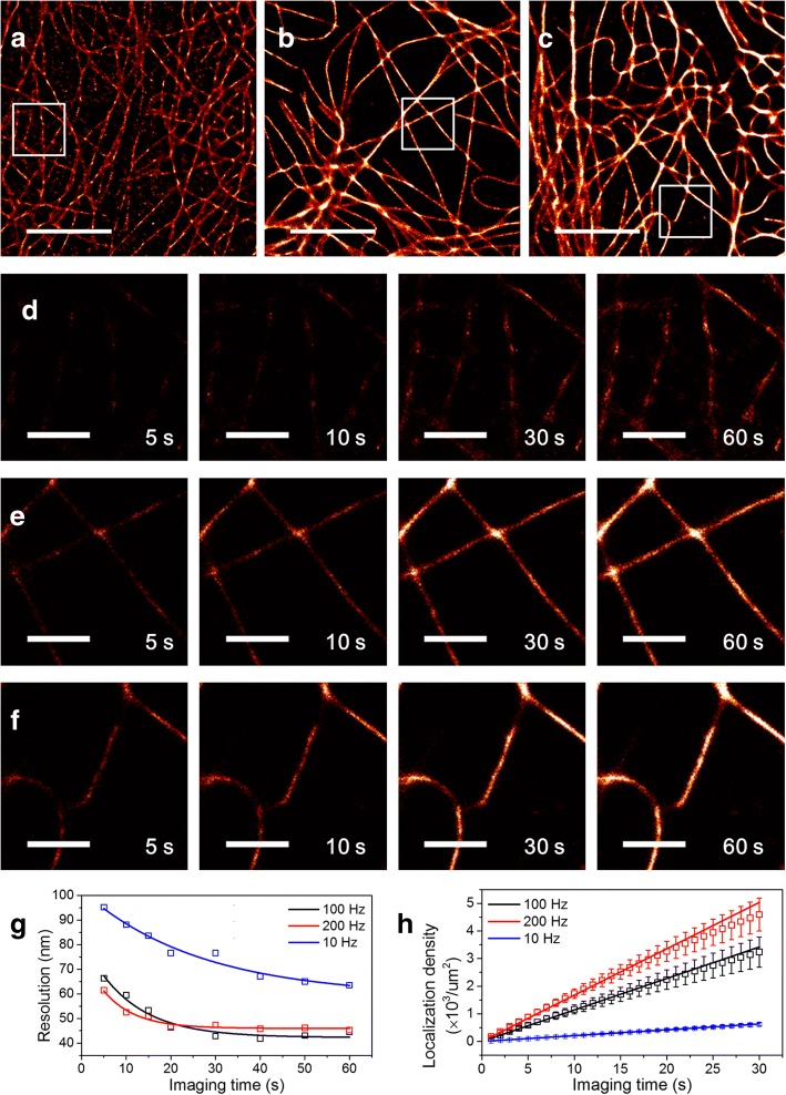
Fig. 3.
| Characterization of the imaging speed of a new microscope. Super-resolution microtubule images of fixed COS-7 cells were used as a model system. (a) The image was reconstructed from 600 frames recorded at a frame rate of 10 Hz with a previous microscope (an EMCCD camera, a long-pass filter, 460 W/cm2 excitation power, 30 nM 9 nt AF488 donor strands, 20 nM 10 nt Cy5 acceptor strands). (b, c) The images were reconstructed from 6000 frames recorded at a frame rate of 100 Hz (b) or 12,000 frames recorded at a frame rate of 200 Hz (c) with a new microscope (an sCMOS camera, a band-pass filter, 460 W/cm2 excitation power, 300 nM 7 nt CF488A donor strands, 300 nM 9 nt Cy5 acceptor strands). An imaging buffer (10 mM Tris-HCl, pH 8.0, 500 mM NaCl, 1 mg/ml glucose oxidase, 5 mg/ml glucose, 0.04 mg/ml catalase, and 1 mM Trolox) was used for all imaging. All images were reconstructed using ThunderSTORM [23] with maximum likelihood fitting method. Total imaging time is 60 s for all images. (d-f) Time-lapse images of the boxed regions in a-c at the specified imaging time. (g) Image resolutions of a-c using Fourier ring correlation method as a function of the imaging time. Open squares indicate measured value and solid lines indicate fitted curves with an exponential decay function. (h) A localization density as a function of the imaging time (100 Hz, black; 200 Hz, red; 10 Hz, blue). The localization density is defined as the number of localization events per um2. To minimize the influence of the region of interest selected for data analysis, the localization density was calculated from 10 different regions of 5 different cells. Squared boxes indicate the average and error bars indicate the standard deviation. The increase rates of the localization density were 21 (10 Hz), 114 (100 Hz), and 168 (200 Hz) localizations/um2/s. We obtained 5.4 times increase for 100 Hz imaging, and 8 times increase for 200 Hz imaging compared to the old microscope. Scale bars: 5 um (a-c), 1 um (d-f)

