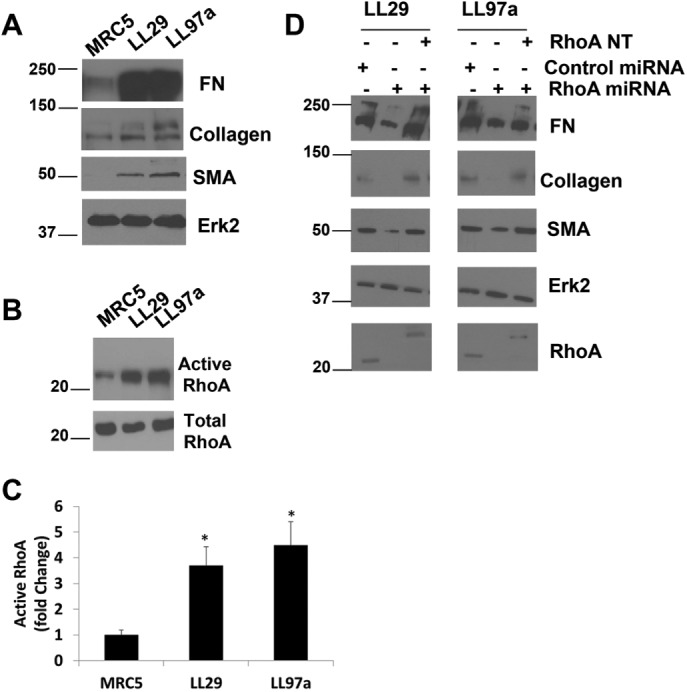
FIGURE 1:

RhoA is required for the IPF phenotype. (A) MRC5, LL29, and LL97a cell lysates were analyzed by Western blotting for FN, collagen I, SMA, and Erk2 (as a loading control). (B) MRC5, LL29, and LL97a cells were lysed, and active RhoA was precipitated from lysates using GST-RBD and immunoblotted with RhoA antibodies. (C) Quantification of RhoA activity in three independent assays. p < 0.05 vs. MRC5 as determined by a t test. (D) LL29 and LL97a cells were infected with an adenoviral miRNA against RhoA for 48 h to knock down RhoA expression. Cell lysates were analyzed by Western blot for expression of RhoA, FN, collagen I, SMA, and Erk2. (D) LL29 and LL97a cells were infected with RhoA miRNA-encoding adenovirus or a control adenovirus for 48 h. After 48 h, cells were transfected with a myc-RhoA NT construct for 24 h, where indicated. After a total of 72 h, total cell lysates were analyzed by Western blot for FN, collagen, SMA, Erk2, and RhoA expression. Note that the position of the myc-RhoA NT construct was detected higher in the blot than the endogenous RhoA.
