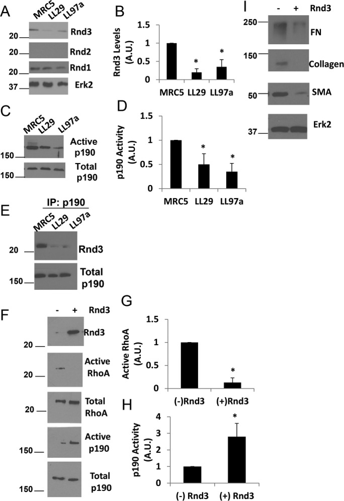
FIGURE 2:

Rnd3 and p190 regulate RhoA activity in IPF cells. (A) MRC5, LL29, and LL97a cell lysates were analyzed by Western blotting for Rnd1, Rnd2, Rnd3, and Erk2 (loading control). (B) Quantification of Rnd3 expression from three independent assays. p < 0.05 vs. MRC5 as determined by a t test. (C) MRC5, LL29, and LL97a cells were lysed and activation of p190 was determined using the GST-RhoAQ63L pull-down assay and immunoblotting with p190 antibodies. (D) Quantification of p190 activity from three independent assays. p < 0.05 vs. MRC5 as determined by a t test. (E) MRC5, LL29, and LL97a cells were lysed in immunoprecipitation buffer and p190 was immunoprecipitated from the cell lysates. Immunoprecipitates were then blotted for the presence of Rnd3. (F) LL29 cells were transfected with Rnd3 cDNA. Cell lysates were then analyzed for RhoA activity through a GST-RBD pull-down assay and p190 activity through a GST-RhoAQ63L pull-down assay. Western blot analysis of pull downs and total cell lysates were analyzed for levels of Rnd3, RhoA, and p190. (G, H) Quantification of RhoA activity (G) and p190 activity (H) from three independent assays. *p < 0.05 vs. (–) Rnd3 as determined by a t test. (I) LL29 cells were transfected with Rnd3 cDNA. Cell lysates were subjected to Western blot analysis for FN, collagen I, and SMA, as well as Erk2 (loading control).
