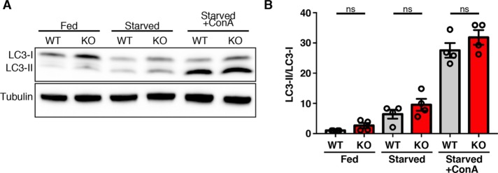
FIGURE 6:
LC3 lipidation and autophagic flux is unimpaired in WDR41 KO cells. (A) Immunoblot analysis of LC3 levels in wild-type and WDR41 knockout cells under fed, starved, and starved cells treated with the v-ATPase inhibitor concanamycin A (ConA). (B) Quantification of LC3-II/tubulin levels (mean ± SEM, n = 4, Sidak’s multiple comparisons test).

