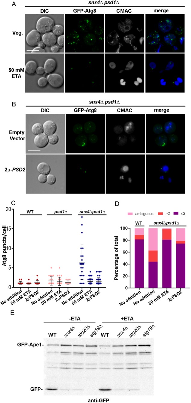
FIGURE 6:

snx4Δpsd1Δ cells are deficient in PE trafficking to the autophagosome resulting in the loss of fusion to the vacuole. (A) Maximum projection micrographs showing snx4Δpsd1Δ cells expressing GFP-Atg8 grown in standard synthetic medium or medium supplemented with 50 mM ethanolamine. Vacuoles were visualized using CMAC. The sale bar indicates 5 μm. (B) Maximum projection micrographs showing snx4Δpsd1Δ cells expressing GFP-Atg8 and overexpressing PSD2 (2μ-PSD2) are shown. The sale bar indicates 5 μm. (C) The number of Atg8 puncta per cell was quantified in wild-type and mutant cells in the absence and presence of 50 mM ethanolamine (ETA), or overexpression of PSD2. Each point is a single cell and the mean and SD are indicated. (D) The number of vacuoles per cell was quantified in wild-type and snx4Δpsd1Δ cells in the absence of 50 mM ETA or 2μ-PSD2 and snx4Δpsd1Δ cells either grown in 50 mM ETA or expressing 2μ-PSD2. The cells were binned into three categories, one or two CMAC-stained vacuoles (a single vacuole may contain multiple lobes that are clearly connected), more than two CMAC-stained vacuoles, or “ambiguous,” which are cells that do not stain well with CMAC, and presented as percentages of the total number of cells quantified. (E) Representative immunoblot analysis of GFP-Ape1 processing in cells grown to log phase in standard or 50 mM ETA-supplemented medium in wild-type or indicated mutant cells. Anti-GFP was used to detect full length GFP-Ape1 and free GFP proteolytic fragment. Note the reduction in GFP-Ape1 processing in snx4Δ and atg20Δ cells compared with wild-type cells, which is still maintained when cells are grown in ETA. The atg19Δ mutant lacks the Ape1 sorting receptor and serves as a control.
