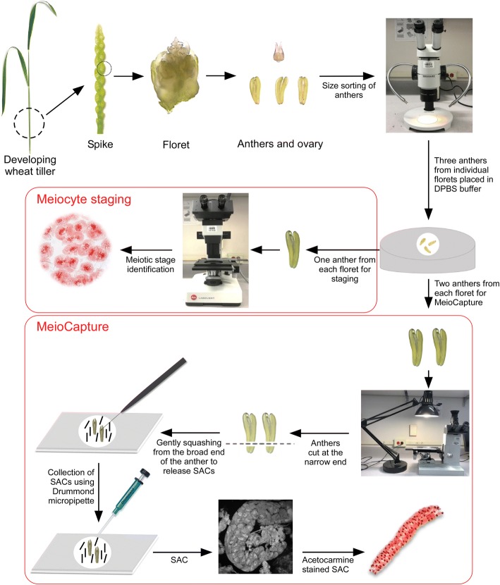
Fig. 2.
Schematic representation of MeioCapture technique for meiocyte isolation in wheat. A developing wheat tiller, young spike, floret, ovary and the anthers isolated from the floret are shown. For staging and meiocyte isolation, anthers are isolated and size sorted under a dissection microscope. After careful staging of one out of the three anthers from each floret, meiocytes are collected from pooled anthers by MeioCapture method. Anthers in DPBS buffer are cut at the narrow end, squeezed gently from the broad end to release the sporogenous archesporial columns (SACs) which are collected using a Drummond microdispenser

